The heart and blood vessels deliver oxygen and nutrients to every tissue in the body, so monitoring cardiovascular health with various medical specialists is invaluable to your overall health. Nuclear medicine uses medical imaging, often with small amounts of radioactive material, called radiotracers, to show how organs and tissues function. Unlike imaging methods that focus mainly on structure, nuclear medicine captures functional information, such as blood flow and metabolic activity. Here’s more information on nuclear screening innovations for monitoring cardiovascular health:
How Nuclear Medicine Supports Diagnosis
During a nuclear medicine exam, a radiologist administers a radiotracer by injection, orally, or by inhalation. The tracer travels to a specific organ or tissue, where it emits a small amount of energy. A specialized camera detects this energy and creates images that reflect how a body area is functioning. Physicians review these images alongside other test results to help inform a diagnosis for various health needs. Nuclear medicine offers several screening options, including:
- Bone Scans: These show areas of increased or decreased bone activity, which may relate to fractures, infections, or other bone conditions.
- Thyroid Uptake Scans: Thyroid scans measure how much tracer the thyroid gland absorbs to assess thyroid function.
- Hepatobiliary Scans: These track bile flow from the liver through the gallbladder and into the small intestine.
- Positron Emission Tomography Scans: These measure metabolic activity, blood flow, and other processes in organs such as the heart and brain.
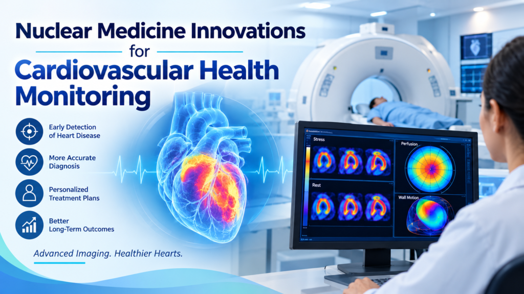
Many of these scans focus on viewing organs outside the chest. For cardiovascular concerns, chest imaging is more common because the heart, major blood vessels, and lungs sit within the chest cavity. Imaging this region lets physicians view the heart muscle and surrounding structures in a single study. Cardiac nuclear exams, such as myocardial perfusion imaging, show how blood moves through the heart muscle at rest and during stress. Some symptoms that appear cardiac originate in the lungs; chest imaging can help physicians distinguish between these sources.
How Imaging Scans Aid Treatment
Computed tomography (CT) scans and X-rays are not nuclear medicine procedures, but they often complement nuclear imaging in cardiovascular care. A radiologist may order imaging tests and nuclear medicine scans to gather more information about someone’s condition. A chest X-ray uses a small dose of radiation to produce a two-dimensional image of the heart, lungs, and blood vessels; physicians use it to assess the heart’s size and shape. An X-ray also helps reveal fluid in the lungs or changes in the major arteries.
A CT scan combines a series of X-ray images taken from different angles. A computer processes these images into detailed cross-sections of the body. Cardiac CT scans can show calcium buildup in the coronary arteries, while CT angiography uses contrast dye to outline the blood vessels. Physicians may use imaging findings to plan procedures, select treatment approaches, and monitor changes after treatment.
How PET Scans Help Cardiovascular Conditions
Cardiac PET scans measure blood flow through the heart muscle and assess tissue viability. During a PET perfusion study, the patient receives a tracer such as rubidium-82 or ammonia N-13. A radiologist takes images at rest and again after the patient receives a medication that simulates exercise. Comparing these sets of images helps medical teams identify areas of the heart that are receiving less blood during stress. PET scans also assess whether heart muscle with reduced blood flow remains metabolically active, guiding decisions about bypass surgery or stent placement.
Get Nuclear Medicine Screenings
Nuclear medicine provides functional information that can support cardiovascular evaluation alongside CT scans and X-rays, and radiologists interpret these studies and work with your cardiologist to coordinate testing. They can explain how each exam works, how to prepare, and what your results mean. If you have questions about cardiovascular imaging, contact a radiologist to learn which screening options may suit your needs.